Encoris Products
Models for Medical Training & Demonstration
Trusted by leading medical device companies, Encoris supports product marketing teams with high-quality, fast-turnaround surgical training models designed to promote implant success and adoption. Orthopedic leaders turn to Encoris for reliable, hands-on training solutions that elevate education and engagement.


Built for Precision & Made to Last
Extremity Models for Medical Training & Demonstration
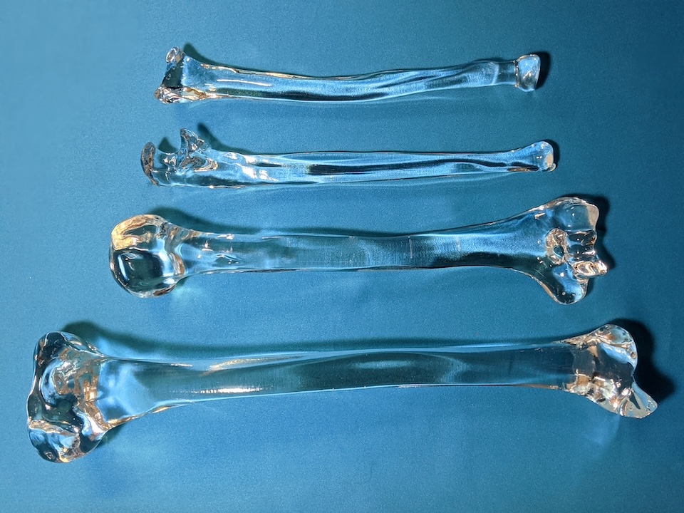
Encoris’ specialized design and manufacturing process streamlines the development of acrylic bone models, no matter how complex the anatomy or implant assembly. We guide clients through each step, often in real time, to ensure every model is accurate from the start and delivered on time with complete confidence.
Our translucent extremity models support clear, in-depth demonstrations of a wide range of orthopedic surgical techniques, including:
Open Reduction/Internal Fixation (ORIF)

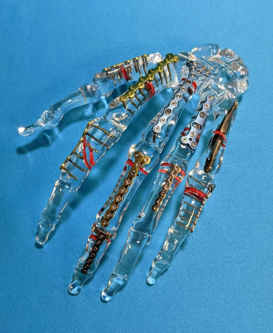
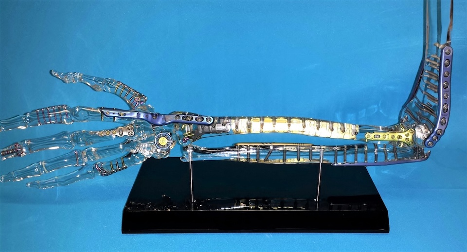
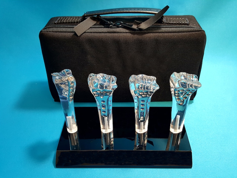
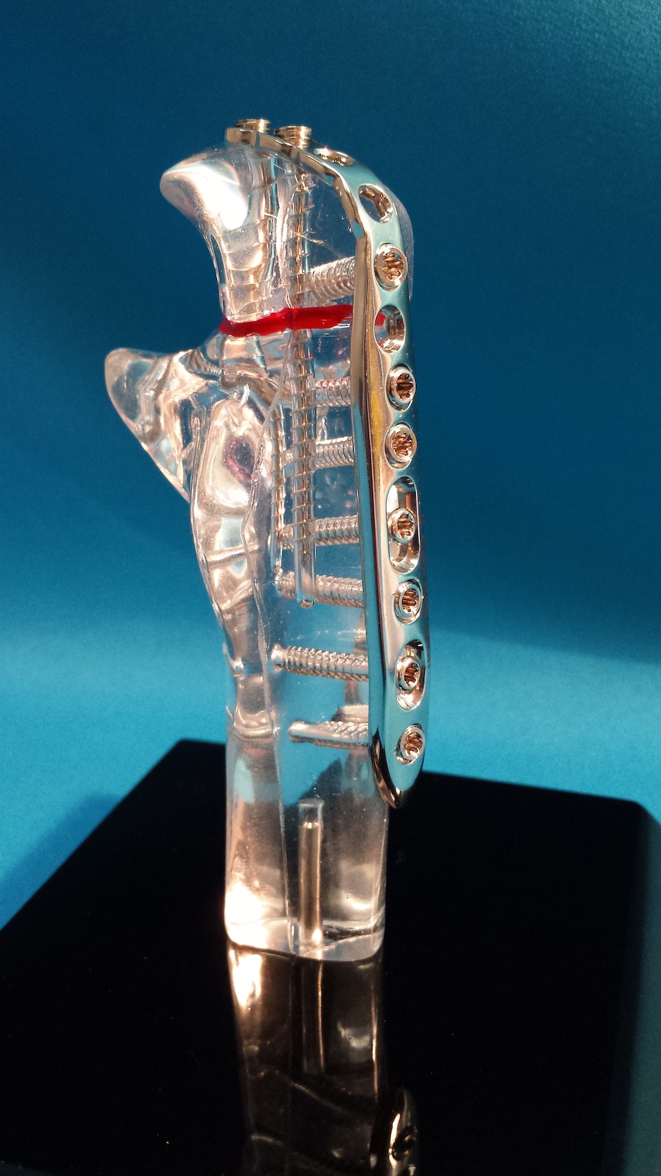
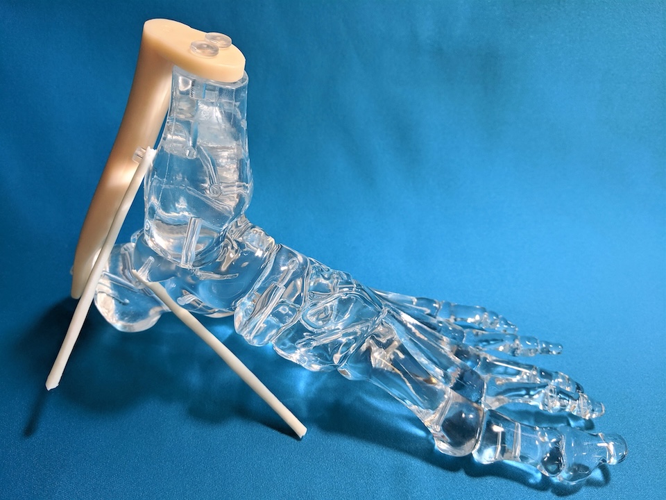
Detailed extremity models help illustrate hardware placement and surgical procedures for internal fixation of displaced, comminuted, or unstable fractures of the long bones, metacarpals, metatarsals, and phalanges. We offer highly accurate models of the humerus, radius, ulna, tibia, fibula, femur, foot, and hand—ideal for training and demonstration, especially when introducing procedures to those with limited prior knowledge.
Percutaneous Pinning
Our models are well-suited for demonstrating wire fixation and percutaneous pinning, particularly useful for procedures involving small bones like the phalanges of the hands and feet. The translucent design allows for clear visualization from every angle, making it easy to showcase both the technique and hardware placement.
Tendon Repair
Patients and even healthcare support staff may have limited understanding of tendon repair or tendon transfer procedures, especially in relation to the underlying bony anatomy. Clear anatomical models make it easier to explain tendon positioning and surgical sites with clarity and precision.
Carpal Tunnel Release
Carpal tunnel syndrome is increasingly common in today’s world of repetitive motion and computer use, leading to a growing need for carpal tunnel release procedures. Our hand models are ideal for illustrating the complexities of nerve entrapment and explaining the surgical approach with clarity.
Engineered to Resist Cracking, Tarnishing & Fading
Durable Orthopedic Joint Models
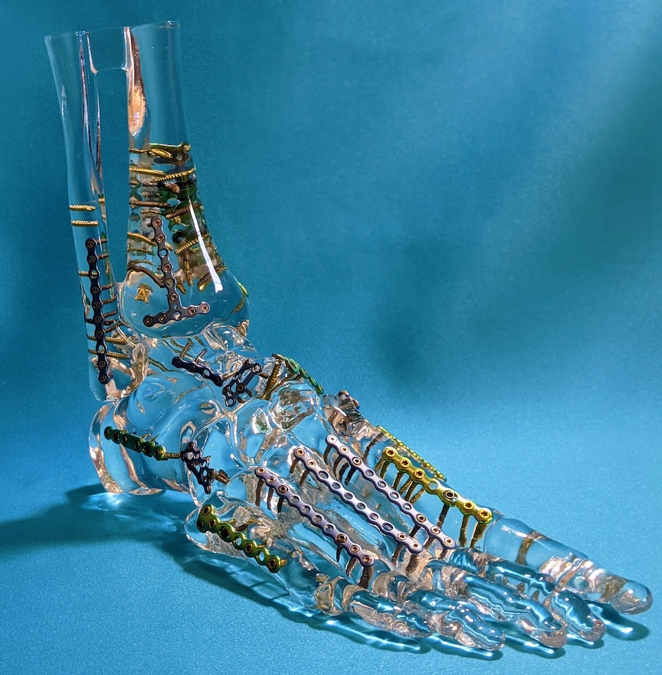
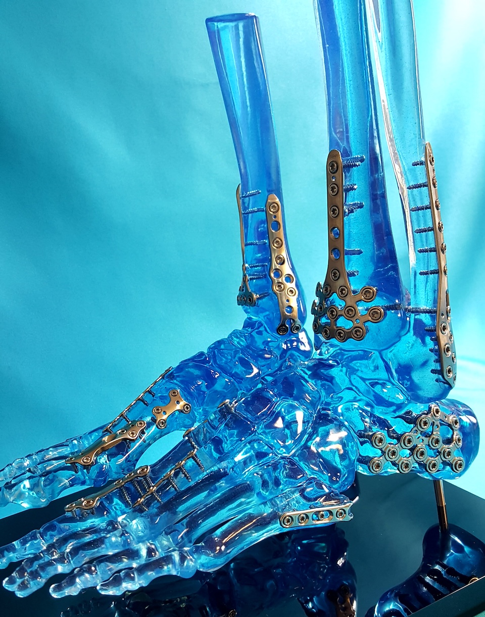
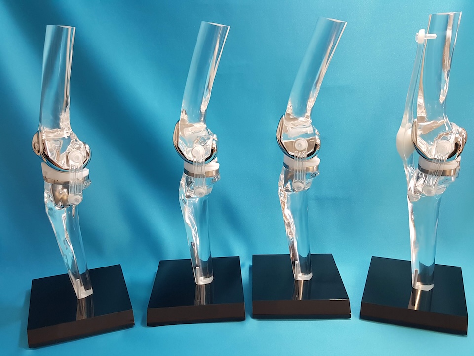
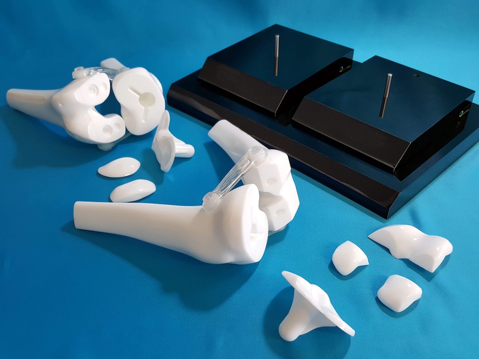
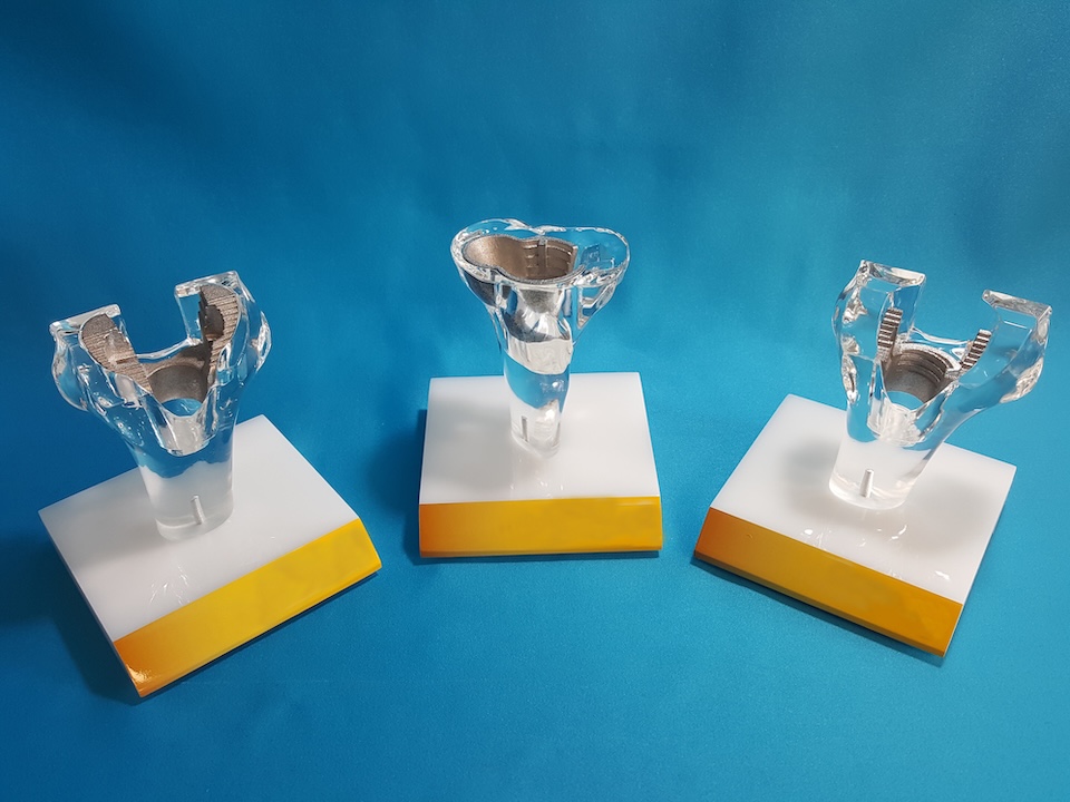
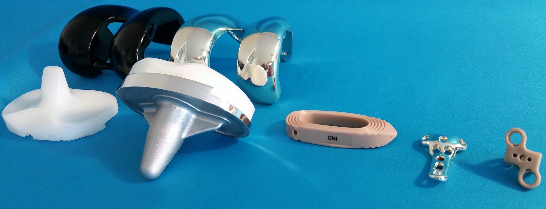
Our joint models do not crack, tarnish, or fade! Demonstrate and conceptualize many procedures, aiding in showing exact hardware and prosthetic positioning and surgical approaches.
First impressions matter. Anatomical joint models designed for surgeon desktop display and patient education help keep your company’s name and products front and center. Encoris joint models are available in a range of clear and solid colors, fully customizable to fit any implant specification. Choose from a variety of support bases, including vibrant printed logos and rechargeable LED lighting options for added impact.
Below is a joint-by-joint overview of some common procedures easily demonstrated using our orthopedic joint models:
Shoulders
- Acromioplasty
- Capsulotomy
- Rotator cuff repair
- Shoulder arthroscopy
- Total shoulder replacement
Wrists
- Carpal tunnel release
- Distal radius fracture fixation
Ankles
- Achilles tendon repair
- Ankle arthrodesis
- Ankle arthroscopy
- Ankle cheilectomy
Elbows
- Elbow arthroscopy
- Lateral epicondyle release
- Reconstructive elbow surgery
Knees
- Anterior cruciate ligament (ACL) repair
- Knee arthroscopy
- Lateral collateral ligament (LCL) repair
- Medial collateral ligament (PCL) repair
- Meniscus repair
- Partial knee replacement
- Plica excision
- Posterior cruciate ligament (PCL) repair
- Total knee replacement
- Unicompartmental knee replacement

Clear, Efficient Implant Demonstrations
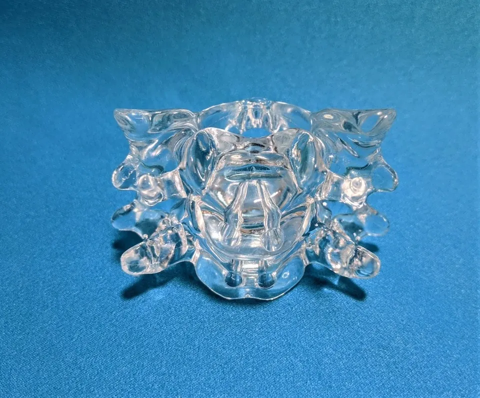
Industry-Leading Spine Models
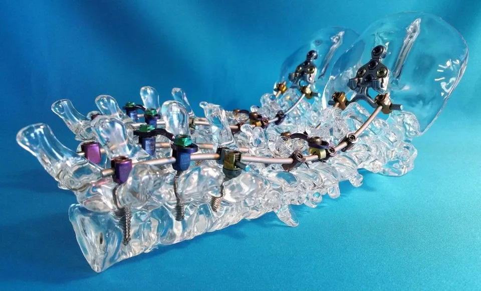
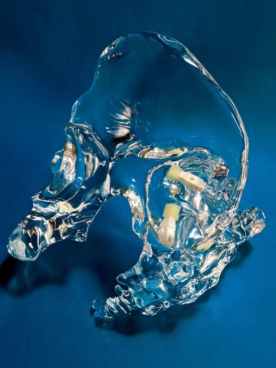
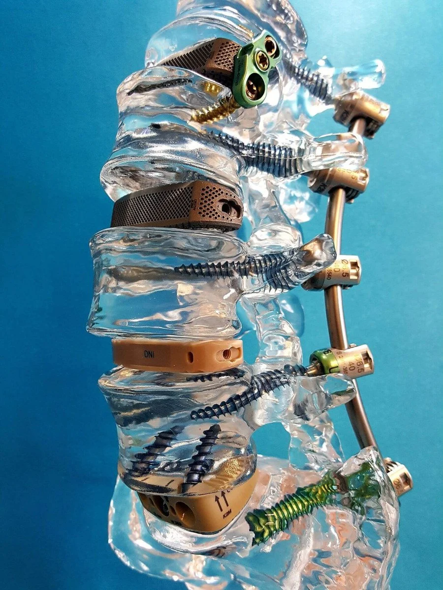
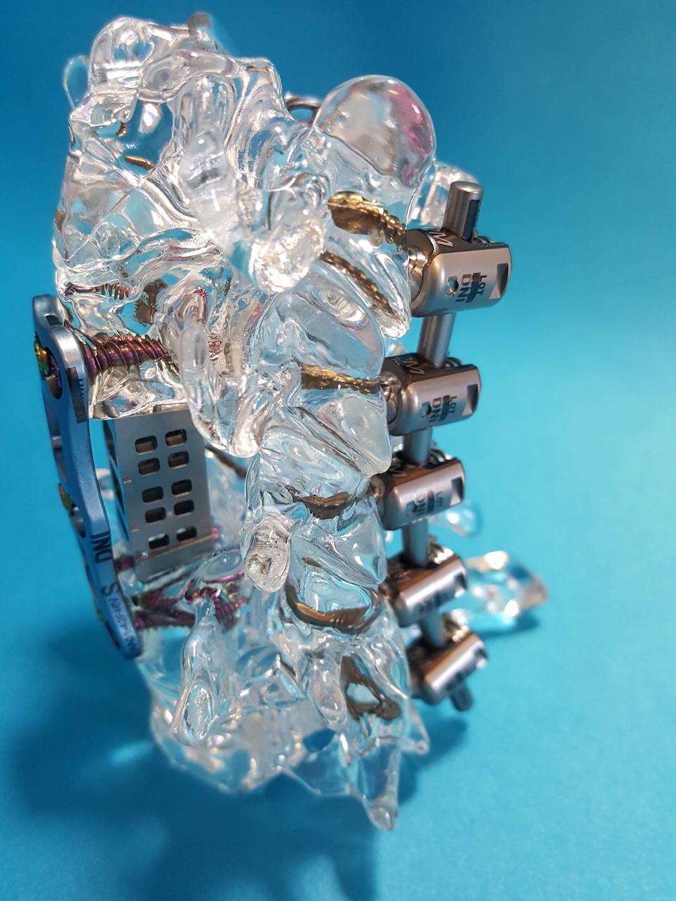
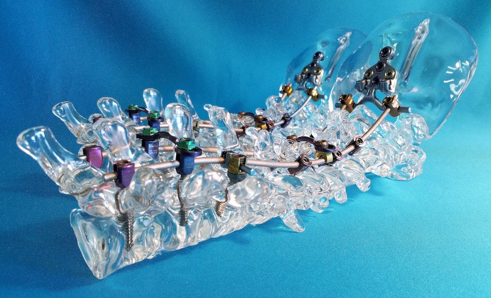
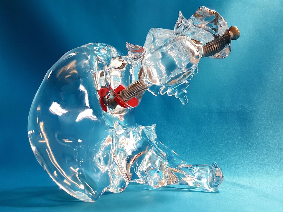
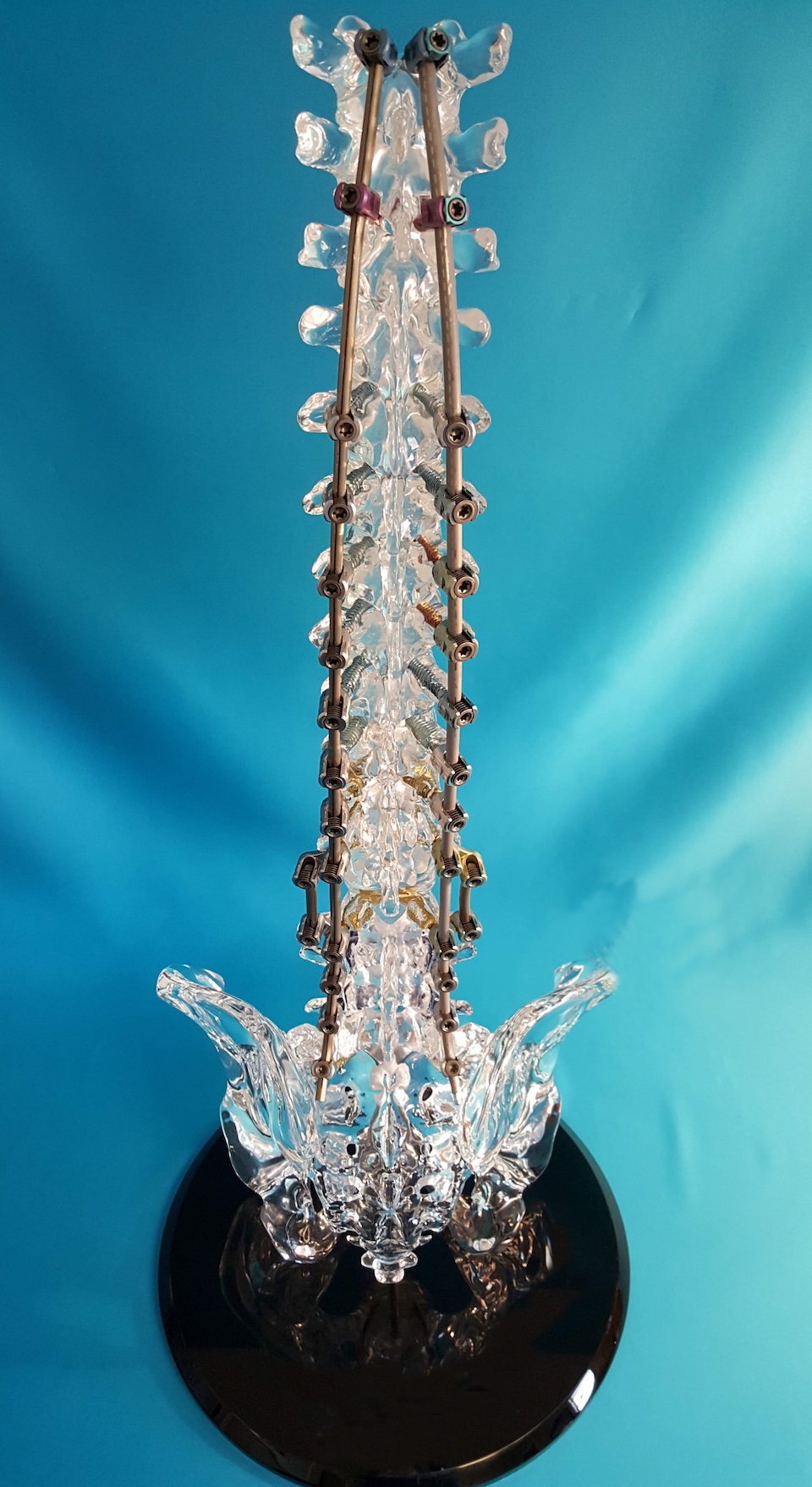
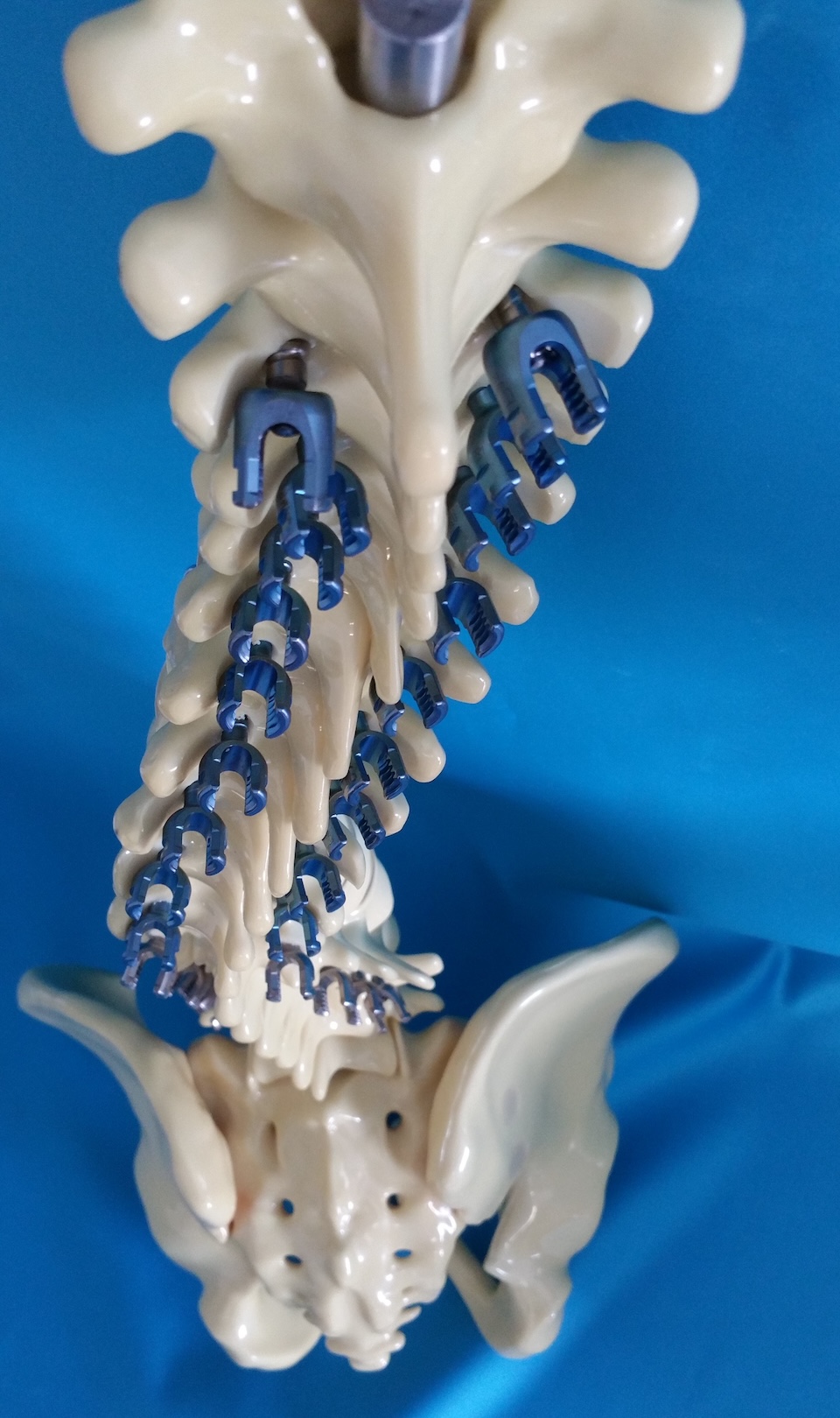
Encoris spine models accurately showcase device details and are ideal for training, sales demonstrations, and as striking display pieces.
Whether it’s a one-level spine model or a complex full acrylic spine, our skilled model makers are equipped to tackle any challenge. Each bone is meticulously crafted and refined, and our proprietary embedding process guarantees implants with brilliant, clear visuals that resist tarnishing, fading, and cracking.
Our comprehensive spinal models can demonstrate a wide range of surgical procedures, including:
- Anterior cervical discectomy and fusion
- Anterior lumbar discectomy
- Anterior lumbar interbody fusion
- Bone grafting
- Cervical fusion
- Discectomy
- Foraminotomy
- Foraminectomy
- Hemilaminectomy
- Intrathecal pump placement
- Kyphoplasty
- Laminectomy
- Laminoplasty
- Laminotomy
- Lumbar fusion
- Microdiscectomy
- Posterior lumbar interbody fusion (PLIF) with cage placement
- Spinal stimulator placement
- Thoracic fusion
- Transforaminal lumbar interbody fusion (TLIF) with cage placement
Setting Standards in Model Making
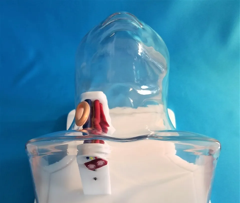
Cardiology & Vascular Models
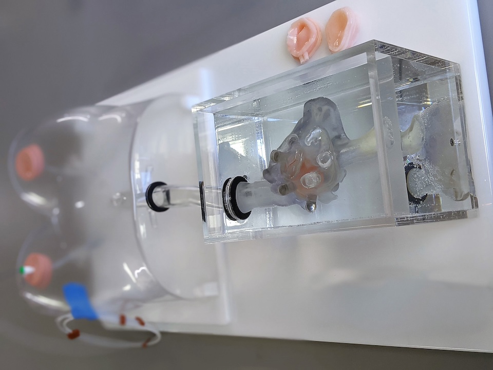
Leveraging our deep understanding of physiology and tactile response, we customize vascular product solutions to help you effectively showcase and demonstrate your medical devices and surgical procedures.
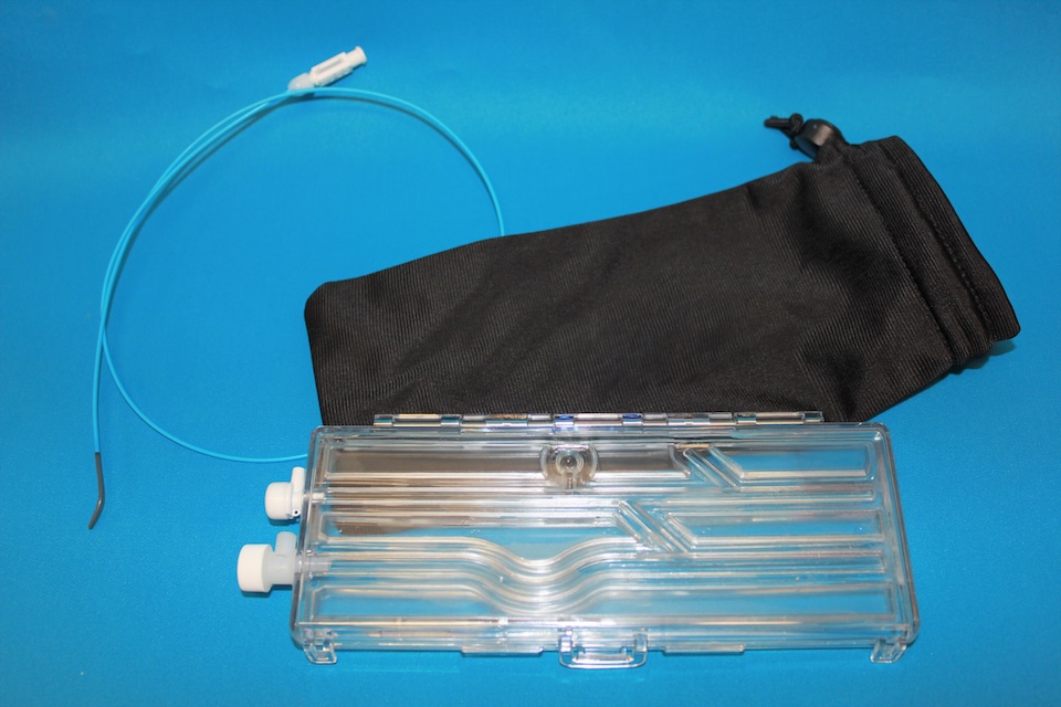
Professional anatomical models are ideal for showcasing medical devices used in vascular surgery, interventional cardiology, interventional radiology, and peripheral vascular procedures. These devices may include stents, catheters, clips, and filters. With a wide range of diagnostic and interventional applications, anatomical models offer a clear and effective way to demonstrate and teach procedures to healthcare professionals across cardiology and vascular specialties.
Encoris is advancing the development of next-generation cardiovascular and orthopedic surgical training models. Designed to complement cadaver use, these models provide a practical alternative for specific types of device training and perioperative or preoperative planning. While cadavers remain essential in many areas of medical education, Encoris models offer repeatability, portability, and a radiation-free environment — making them an efficient solution for modern training needs.
Examples of procedures demonstrated with these models
- Angiography
- Angioplasty
- Atherectomy
- Bypass for peripheral artery disease (PAD)
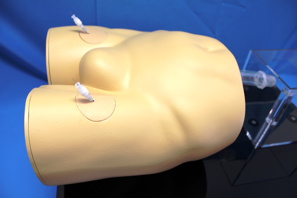
- Dialysis access creation and maintenance
- Life-like, ultra-sound responsive skin
- Embolization
- Stent placement
- Vein grafting
- Venous stripping
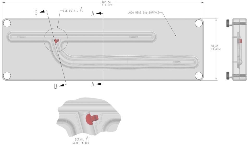
Customizable and flexible orthopedic models
FlexBones Orthopedic Models
Customizable and flexible, FlexBone orthopedic models offer a clear advantage by functioning as both static displays and interactive demonstration tools.
FlexBones are innovative, cutting-edge alternatives to traditional orthopedic models, designed to simplify medical sales and patient education. They can be used as static displays or interactive demo models to showcase implant function and anatomical movement.
How do they compare to acrylic models?
FlexBones are virtually unbreakable. Their flexible design eliminates concerns about damage during handling, transport, or demonstration. FlexBones can be bent, manipulated, and withstand repeated use — always returning to their original shape. After demonstrating and removing an implant, the same pathway can be reused. FlexBones deliver unmatched durability, clarity, and reliability in orthopedic modeling.
Can they be customized?
FlexBones can be customized to fit your specific implant needs and are available with pathology features, making them ideal for teaching and device demonstrations.
What is the price point?
FlexBones cost 30–40% less than acrylic models, offering an attractive, versatile, and durable solution for field sales, education, and patient engagement.
Better Practice. Better Doctor. Better Care.
With our solutions, every learner can practice until ready. Every educator can teach with confidence. Every medical device can be seen, touched, and understood and every patient can trust.
Newsletter Signup
Stay up-to-date with Science Care, medical research, and health and wellness tips.